Plant Leaf Cell Under Microscope Labeled - Proliferation Of C Boidinii On Growing A Thaliana Leaves A Download Scientific Diagram : First find water plant cells using 4x objective, then change to 10x and focus and draw, then turn to 40x and draw.
byZackary Gambale-0
Plant Leaf Cell Under Microscope Labeled - Proliferation Of C Boidinii On Growing A Thaliana Leaves A Download Scientific Diagram : First find water plant cells using 4x objective, then change to 10x and focus and draw, then turn to 40x and draw.. When viewed under the microscope, it's possible to see the epidermal cells that tend to be irregular. Cover leaf with a cover slip (small square) 2. Stomata under microscope labeled written by macpride sunday, november 4, 2018 add comment edit. Animal cell under microscope labeled. We have sets that will allow your students to study and identify the similarities and differences between monocot and dicot tissues and structures.
The plant cell is the basic structural and functional unit found in the members of the kingdom plantae. Microscope, the structures to be labeled are indicated in bold type than minutes. Learn even more about plants by studying different sections of real leaves. All you need is a fresh leaf specimen (use one without many holes or blemishes), a plain glass. View under the microscope and sketch the cells at each magnification.
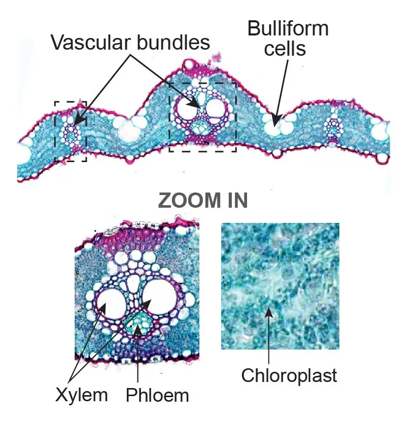
Plant Tissue Under A Microscope Xylem And Phloem Rs Science from rsscience.com Label the cells as they appear under high power. Label the xylem and phloem Cell is a tiny structure and functional unit of a living organism containing various parts known as organelles. Animal cell under microscope labeled. Under the microscope have students determine the field diameter of the compound microscope objectives. The numerous green chloroplasts allow the cell to. Learn the structure of animal cell and plant cell under light microscope. Plants capture the sun's light within their green leaves.
Make sure the leaf does not fold…it must remain flat!
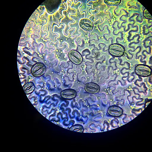
A) collect a suitable leaf and all other materials. Magnifying and observing cells bioed online. Elodea leaf cells with structures labeled chloroplasts and mitochondria move within elodea leaf cells; An elodea leaf was mounted in pondwater between a slide and coverslip with a silicon spacer. In addition to the epidermal cells, one will also see the leaf spores (stomata) in between the epidermal cells. Animal cell under microscope labeled. Inside a leaf's cells are green organelles — chloroplasts — which do all this hard work of producing the food that feeds the plant… and, in fact, the whole rest of the world, too! Inside a leaf's cells are green. Place the slide under the microscope. Labelled diagram of a plant cell under microscope posted on march 18 2011 by admin onion cells stained with methylene blue look at the images of onion cells as they would be seen under a microscope draw each magnification label appear high picture plant and animal cell diagrams vs science for kids. In addition to guard cells, it is also possible to identify pavement cells around the guard cells. Elodea leaf cell under microscope labeled written by macpride tuesday, august 20, 2019 add comment edit. Leaf structure under the microscope.
Add a drop of salt solution (hypertonic solution) to the side of the coverslip and observe the. Elodea leaf cells with structures labeled chloroplasts and mitochondria move within elodea leaf cells; Elodea are common freshwater aquarium plants. Pick off an entire healthy looking elodea leaf, with fingers or small scissors and place it on the microscope slide. Under the microscope have students determine the field diameter of the compound microscope objectives.
Leaf Structure Under The Microscope from www.microscopemaster.com View under the microscope and sketch the cells at each magnification. If you are trying out a new leaf type, be sure to test it yourself before doing it with students). Label the cells as they appear under high power. In addition to guard cells, it is also possible to identify pavement cells around the guard cells. Organelles (chloroplasts and mitochondria) move within elodea densa leaf cells. Pick off an entire healthy looking elodea leaf, with fingers or small scissors and place it on the microscope slide. Although some botanists divide this category into several species. Generalized cell is used for structure of animal cell and plant cell to present the.
Microscope, the structures to be labeled are indicated in bold type than minutes.
The numerous green chloroplasts allow the cell to. When viewed under the microscope, it's possible to see the epidermal cells that tend to be irregular. Place the tip of water plant leaf in the water. Piefa video using water sustainably through science. Learn the structure of animal cell and plant cell under light microscope. Animal cell under microscope labeled. Label the xylem and phloem If you are trying out a new leaf type, be sure to test it yourself before doing it with students). You can make your own microscope slide of a leaf section and view it under high power with a compound microscope to see cell detail. Magnifying and observing cells bioed online. View a prepared slide of elodea (anacharis), which is an aquarium plant. Elodea leaf cell under microscope plant cell biology science. If the student can see only a thick, dark mass when looking under the microscope, the slice is too thick.
Label the cells as they appear under high power. Rigid walls typically made of cellulose surround plant cells. Return to leaf structure under the microscope Also indicate the estimated cell size in micrometers under your drawing. Piefa video using water sustainably through science.
Here S How Plant And Animal Cells Are Different Howstuffworks from media.hswstatic.com Pick off an entire healthy looking elodea leaf, with fingers or small scissors and place it on the microscope slide. Add a drop of water (hypotonic solution) and a coverslip and observe the chloroplasts (green structures) and the cell walls. Typically, the stomata are bean shaped and will appear denser (darker) under the microscope. Label a microscope slide b) bend the leaf to break the surface or tear the leaf from the edge c) tear off some epidermis, the transparent thin layer of surface cells d) cut the epidermal layer from the leaf, place on a microscope slide Hydrilla verticillatea leaf under the microscope hydrilla (esthwaite waterweed, waterthyme or hydrilla) is a genus of aquatic plant that is usually treated as containing only one species: Organelles (chloroplasts and mitochondria) move within elodea densa leaf cells. An elodea leaf was mounted in pondwater between a slide and coverslip with a silicon spacer. Generalized cell is used for structure of animal cell and plant cell to present the.
Animal cell under microscope labeled.
However, any broad flat leaf should work. Label the xylem and phloem Plants capture the sun's light within their green leaves. Actin based photo orientation movement of chloroplasts in plant. Place the tip of water plant leaf in the water. Piefa video using water sustainably through science. Plant guard cells with stoma fully labeled 85064282 image. Also indicate the estimated cell size in micrometers under your drawing. Elodea are common freshwater aquarium plants. Elodea are common freshwater aquarium plants. When viewed under the microscope, it's possible to see the epidermal cells that tend to be irregular. Leaf stomata under microscope labeled › stomata under microscope labeled. Rigid walls typically made of cellulose surround plant cells.
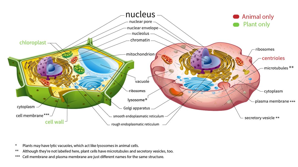
Inside a leaf's cells are green organelles — chloroplasts — which do all this hard work of producing the food that feeds the plant… and, in fact, the whole rest of the world, too! plant cell microscope labeled. On this page, we will learn about what is a plant cell, definition, structure, model, labeled plant cell diagram, its cell organelles and the difference between plant cell and animal cell.

Post a Comment