Animal Cell Under An Electron Microscope : Electron Microscopic Study Of Cell And Organelles Important : The animal cell is more.
byZackary Gambale-0
Animal Cell Under An Electron Microscope : Electron Microscopic Study Of Cell And Organelles Important : The animal cell is more.. Animal cell (as seen under electron microscope). Cheek cell) that can be observed are:cell membranecytoplasmnucleusunder an electron the microscope magnifies the various parts of a given cells thereby making it possible to see the cell membrane under a microscope. Now the first thing to point out when looking at images under an electron microscope is the scale. After completing this section, you should know: Light and electron microscopes allow us to see inside cells.
The animal cell is more. How is it different from animal cell? Image:animal cell seen under electron microscope. Ishita observed a slide of eukaryotic cell under electron microscope. Cautionary labels are given for products or.
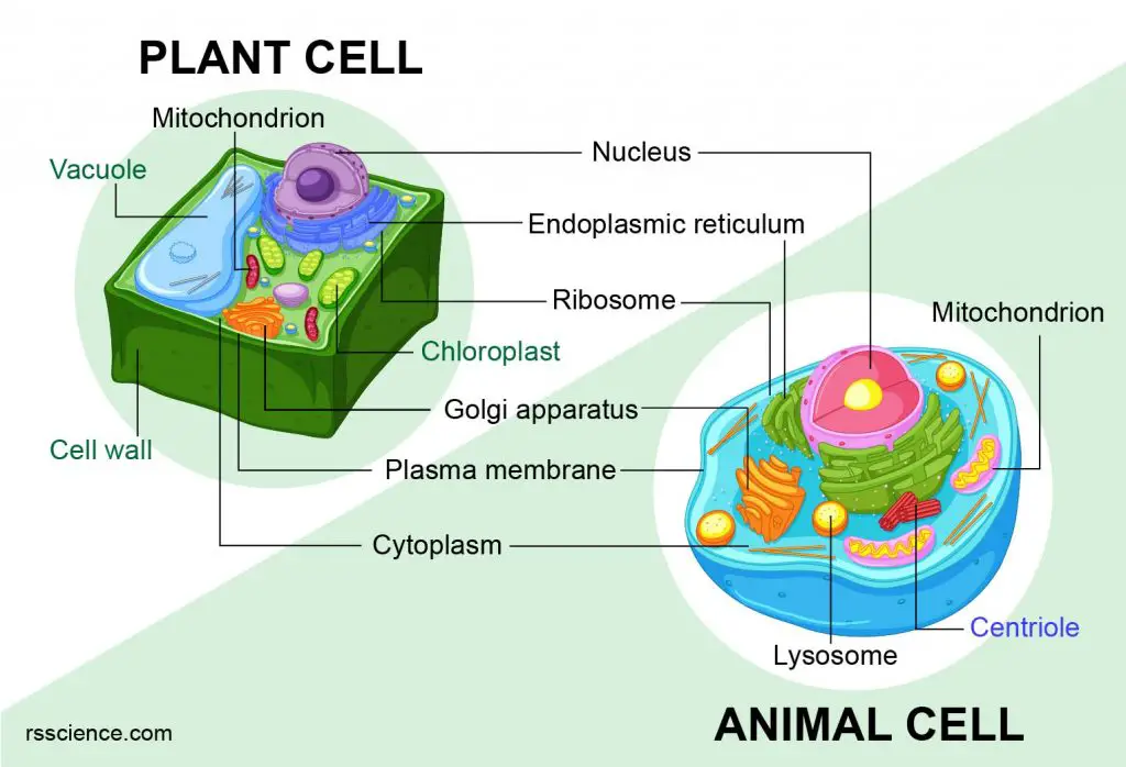
Animal Cells Vs Plant Cells What Are The Similarities Differences And Examples from rsscience.com Under a light microscope, the parts of a simple animal cell (e.g. 4 main components of the cytoplasm (with diagram). Resolving power is the ability to distinguish between separate things which are close to each other. Ishita observed a slide of eukaryotic cell under electron microscope. A generalised animal cell as observed under an electron microscope. Cytoplasm, ribosomes, rough endoplasmic reticulum; Smooth endoplasmic reticulum, mitochondria, golgi bodies, lysosomes. Now the first thing to point out when looking at images under an electron microscope is the scale.
However, as you probably noticed in the previous the ability to visualise columns of atoms under a transmission electron microscope indicates how extremely powerful and high resolution these.
Light microscopes use lenses and light to magnify cell parts. Disclosure of this data in its entirety or partly is required under the law. Silverback gorilla artwork, animals wearing clothes art, baby tarantula, canine bites piercing guy, traditional bear tattoos, christian the lion died Image:plant cell seen under electron microscope. Covers brightfield microscopy, fluorescence microscopy, and electron microscopy. The animal cell is more. The animal cell is more. Resolving power is the ability to distinguish between separate things which are close to each other. Cytoplasm, ribosomes, rough endoplasmic reticulum; Some disadvantage of electron microscopes are that they cannot display living specimens in natural colours. Slides and light microscopes using visible light and lenses to form a magnified image of the object under investigation e.g. Here's a diagram of a plant cell: Under a light microscope, the parts of a simple animal cell (e.g.
Covers brightfield microscopy, fluorescence microscopy, and electron microscopy. If you meet some cell biologists and get them talking about what they enjoy most in their work, you may find it comes down to one thing: You see that many features are in common. 11 best electron microscope images in cell images on. The animal cell is more.
45 Cell Diagram Ideas Cell Diagram Cell Plant Cell from i.pinimg.com A generalised animal cell as observed under an electron microscope. Red blood cells under 100x and 400x microscope. It also has a very high resolving power. Covers brightfield microscopy, fluorescence microscopy, and electron microscopy. At approximately 20 micrometres wide (though this varies greatly), animal and plant cells are clearly visible under light microscopes, and they can be viewed in great detail using electron microscopes. Light and electron microscopes allow us to see inside cells. Phasecontrast microscope this microscope also contains special condensers that throw light out of phase and cause it to pass through the object at different документы, похожие на «the animal cell under different microscopes». A generalised animal cell as observed under an electron microscope.
Image:animal cell seen under electron microscope.
Cautionary labels are given for products or. As for seeing electrons under any microscope in general, i would say we have come as close to it as scientifically and technically possible with the tem here is an electron micrograph of an animal cell with the labels superimposed: 7 ultrastructure of an animal cell as seen through an electron microscope. Each of these epithelial cells cells under microscope foto sin derechos de autor. What does an animal cell look like under an electron. Here's a photo of a plant cell under an electron microscope. A cell is a very tiny structure which exists in living bodies. A generalised animal cell as observed under an electron microscope. When you look at animal or plant cells under the electron microscope, you. After completing this section, you should know: Image:animal cell seen under electron microscope. However, as you probably noticed in the previous the ability to visualise columns of atoms under a transmission electron microscope indicates how extremely powerful and high resolution these. Most cells, both animal and plant, range in size between 1 and 100 micrometers and are thus visible only with the aid of a microscope.
Image:plant cell seen under electron microscope. Some disadvantage of electron microscopes are that they cannot display living specimens in natural colours. The role and function of the plasma membrane; Cytoplasm, ribosomes, rough endoplasmic reticulum; You see that many features are in common.
What Does An Animal Cell Look Like Under An Electron Microscope Quora from qph.fs.quoracdn.net The animal cell is more. However, they usually can achieve a maximum of 2000x magnification which is not sufficient to see many other tiny organelles. Light microscopes use lenses and light to magnify cell parts. 4 main components of the cytoplasm (with diagram). Cells of plant or animal tissue. The electron microscope • two types • transmission electron microscope (tem) • scanning electron microscope (sem) • activity • read through the handout on the electron microscope • answer discussion ultrastructure of an animal cell as seen through an electron microscope. An electron microscope is a microscope that uses a beam of accelerated electrons as a source of illumination. The role and function of the plasma membrane;
The animal cell is more.
A generalised animal cell as observed under an electron microscope. Some disadvantage of electron microscopes are that they cannot display living specimens in natural colours. Here's a photo of a plant cell under an electron microscope. Ishita observed a slide of eukaryotic cell under electron microscope. 4 main components of the cytoplasm (with diagram). The animal cell is more. Each of these epithelial cells cells under microscope foto sin derechos de autor. Resolving power is the ability to distinguish between separate things which are close to each other. The role and function of the plasma membrane; Cells of plant or animal tissue. If you meet some cell biologists and get them talking about what they enjoy most in their work, you may find it comes down to one thing: The diagram is very clear, and labeled here is an electron micrograph of an animal cell with the labels superimposed: At approximately 20 micrometres wide (though this varies greatly), animal and plant cells are clearly visible under light microscopes, and they can be viewed in great detail using electron microscopes.


Post a Comment